Welcome to Vascular Surgery Associates
Providing outstanding clinical care to patients in the greater Southern California area since 1963.
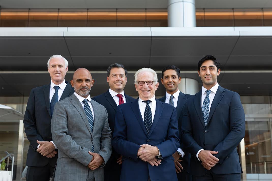
Vascular Surgery Associates (VSA) has been providing outstanding clinical care to patients in the greater Southern California area since the group was established in 1963. Our physicians have extensive experience in all areas of vascular surgery, as one of the busiest practices in the western United States. The physicians of VSA have a long tradition of training future surgeons and reporting our experience with many procedures in peer-reviewed journals.
Articles on carotid artery reconstructions, open abdominal aortic aneurysm surgery, bypasses of leg arteries and treatment of varicose veins have raised the bar for success in these areas. In addition to setting the standard for the traditional treatments of vascular surgical problems, the group has been active in newer, minimally invasive treatments of abdominal aortic aneurysms, peripheral artery blockages and venous disease. Our current involvement in multi-institutional clinical research, such as gene therapy for the treatment of artery blockages in the legs, promises to offer our patients safer, better and less painful solutions to their vascular problems in the future.